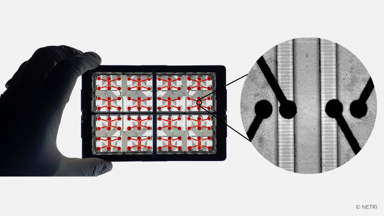
NETRI’s microfluidic devices capabilities.

An example of the experiments you can perform with our microfluidic devices that ensure reproducibility, reliability and ease of use.

1. CC & C - BM1
Co-culture & compartmentalization

Culture of different cell types
Fluidic isolation
Minimal leakage

Explore
2 - HIPSC - BM2
hiPSC culture

Approved alternative methods
7 cell culture & co-culture protocols
2d & 3D cell culture protocols

Explore
3 - 3D CC - BM3
3D-cell culture

Cerebral organoids
Up-to 4 mm 3D-cell culture
Specific open well architecture

Explore
5 - GK-BM5
Growth kinetics & Axotomy

Neurite outgrowth
Specific triangular architecture
Cloudbased software analytics

Explore
6 - SI - BM6
Synaptic isolation

Synaptic compartment
Specific assymetric architecture
Functional analysis

Explore
7 - AT - BM7
Axonal transport

Viruses or proteins
Retrograde or anterograde
Acute or chronic propagation

Explore

Co-culture & Compartmentalization.

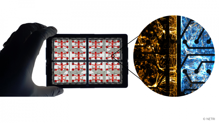
At NETRI we developed compartmentalized microfluidic devices by microchannels technology or membrane technology that create a compartmentalized microenvironments, which allows:

  • Application of different culture media
  • Ensures appropriate differentiation and maintenance of cell cultures
  • Co-culture of different cell types : neurons and neurons or neurons and non-neural cells

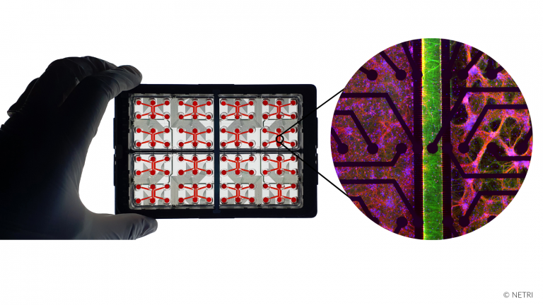
In our NeuroFluidics™ and NeuroFluidics™ MEA line, compartmentalization is provided by microchannels with considerably reduced fluid leakage, compared to the two-channel device, divided by 2 to 3, which ensure:

  • Isolation of neurites and cell bodies to test compounds only on neurites or somas
  • Physiological cellular connection between neurites and non-neuronal cells, as in Innervated skin
  • Modeling of compound mode of action

hiPSC Culture.

Microfluidic coupled with Human induces Pluripotent Stem Cells (hiPSCs) are the most relevant standard for translational research. hiPSC and microfuidic systems are approved alternative methods for discovery and pre-clinical regulatory submission

Our microfluidic devices enable human cells to be cultivate and maintained for up to 3 months in-vitro:

  • 7 cell culture & co-culture protocols of hiPSC & primary cells
  • 2d & 3D cell types protocols
  • Possible cell types: CNS/PNS, muscle, cardiac, gut/liver/kidney organoids, skin epithelia & more
  • Expected differenciated morphology
  • Expression of specific markers and functional activity
  • Development of standard cell culture protocols of any new cell line in 1.5 month

3D-Cell Culture.

Cerebral organoids are composed of several types of human neuronal cells, mainly astrocytes, oligodendrocytes and neurons, which self-organize in 3D. These three-dimensional multicellular structures create cellular interactions and maturation relevant to human pathophysiology. However, despite their relevance, they are currently complex models to implement with significant variability in terms of maturation, morphology and size. To reduce this variability, we have developed at NETRI a unique open well microfluidic devices architecture, that allow to :

  • Grow organoids up to 500 µm before seeding
  • Perfuse growth media for organoids to grow as big as 4 mm in diameter
  • Control the flow of growth media
  • Co-culture of brain organoid and blood-brain barrier cells

NETRI's microfluidic devices would allow not only to co-culture cerebral organoid and other cells but also to record the functional activity of the organoid.

Functional Activity.

Multi Electrodes Arrays (MEA) compatibility approach enables continuous electrophysiological recordings providing data to evaluate neuronal functional activity. With Axion Biosystems, we have developped MEA-capable compartmentalized microfluidic devices which allow investigate a compound’s impact on :

  • Functional activity and recovery
  • Synapse formation
  • Network dynamics

A network can be monitored within all types of NeuroFluidics MEA line microfluidic devices. Electrodes are underneath compartments and microchannels. They are in direct contact with neurons (soma in channels and neurites in microchannels), which allow:

  • Constant monitoring of an entire network while applying cells or compounds on one compartment only
  • Recording spontaneous and induced action potential propagation velocity
  • Couple a functional analysis to a phenotypical analysis
  • Impact of non-neural cells on neurons’s functional activity

Growth Kinetics & Axotomy.

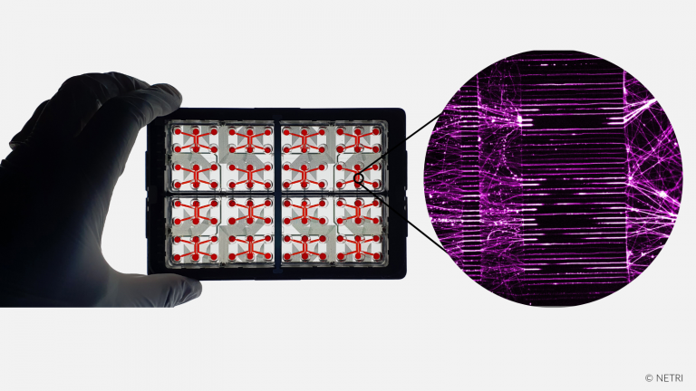
Growth kinetics can be assessed by the triangular design of our DuaLink Delta Ultra. This specific triangular architecture allows the accurate monitoring of neurite growth kinetics in a neuronal culture thanks to the different lenght of microchannels:

Combining the study of axonal growth and compartmentalization, our microfluidic devices allow to perform an axotomy. They also allow to quantify the degeneration of neurons according to molecules, peptides, or factors. NETRI has developed a reproducible protocol to induce localized axonal injury :

  • Axonal injury being achieved only in Channel 2
  • Cells are viable (with/without axonal injury) until day 47 with degeneration of neurites after triton application

NETRI has developed a proprietary software that allows to quantify the axonal growth of neurites according to the application of a pharmacological compound:

  • Automatic extrapolation of Microchannels Grooves in ImagJ
  • Semi-automatic measure of length of Microchannels Grooves

Synapse Isolation.

Asymmetric architecture of DuaLink Shift microfluidic devices allow the reconstitution of a neural network between two neural populations connected through a fluidically isolated synaptic compartment. Thanks to the asymmetry of the connecting microchannels, axons from one population connect to dendrites from the other, with a high probability, only in the middle compartment. The use of multiplexed microchannels offers also the possibility to study subcellular trafficking while altering synapses.

Coupled to MEA, the DuaLink Shift MEA can be used to monitor the electrophysiological impact of a compound when applied in the synaptic compartment only. Alternatively, by seeding cells in the middle compartment when a neural network is matured, interactions between several type of cells and synapses can be monitored.

  • Functional activity and recovery

Axonal transport.

Compartmentalized devices of the NeuroFluidics™ line allow to fluidically isolate two or more cellular populations, even when connected by microchannels. When using neurons, geometrical constraints due to microchannels allows neurites to grow and create functional compartment with distal neurons, both in a unidirectional or bidirectional way. Thanks to the fluidic isolation between compartments, a virus or protein can be applied to one cell population only and their transport can be monitored on the remote compartment or within the axons themselves:

  • Acute or chronic perfusion
  • Retrograde or anterograde transport

Microchannels geometries can also be adapted to create gradients of secreted molecules or to allow migrating cells to pass from one compartment to the other.